

Why Are Filters Overlaid With Xã¢â‚¬â€˜ray Film When Screening A Cdna Library?
ten.seven: Brand and Screen a cDNA Library
- Page ID
- 42823
The commencement step in making a cDNA library is to isolate cellular mRNA. This mRNA extract should correspond all of the transcripts in the cells at the time of isolation, or the cell's transcriptome. This term is used by illustration to genome. However, a genome is all of the genetic information of an organism. In dissimilarity, a transcriptome (usually eukaryotic) reflects all of the genes expressed in a given cell type at a moment in time. Reversetranscribed cDNAs from an mRNA excerpt are as well referred to as a transcriptome…, and likewise, a cDNA library. A cDNA library is a tube full of bacterial cells that have taken up (i.e., been transformed with) plasmids recombined with cDNAs. cDNA libraries fabricated from mRNAs taken from dissimilar cell types or the aforementioned cells grown under dissimilar weather are in consequence, unlike transcriptomes. Each reflects mRNAs transcribed in cells at the moment of their extraction. When cells in a cDNA library are spread out on a nutrient agar petri dish, each jail cell grows into a colony of cells; each cell in the colony is a clone of a starting cell. cDNA libraries can be used isolate and sequence the Deoxyribonucleic acid encoding a polypeptide that you are studying.
Recall that the mature mRNA in eukaryotic cells has been spliced. This means that cDNAs from eukaryotic cells do not include introns. Introns, as well as sequences of enhancers and other regulatory elements in and surrounding a factor must be studied in genomic libraries, to be discussed later. Here we wait at how to brand a cDNA library.
A. cDNA Construction
mRNA is only a few percent of a eukaryotic cell; most is rRNA. But that small corporeality of mRNA tin can be separated from other cellular RNAs by virtue of their 3' poly(A) tails. Simply pass a total RNA extract over an oligo-d(T) column (illustrated below).
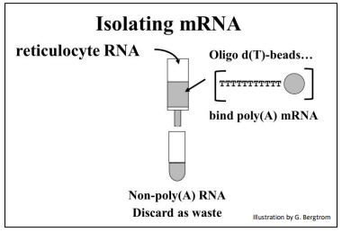
The strings of thymidine (T) can H-bail with the poly(A) tails of mRNAs, tethering them to the column. All RNAs without a 3' poly(A) tail volition period through the column as waste matter. A second buffer is passed over the column to destabilize the A-T H-bonds to permit elution of an mRNA fraction. When free' oligo d(T) is added to the eluted mRNA, it forms H-bonds with the poly(A) tails of the mRNAs, serving as a primer for the synthesis of cDNA copies of the poly(A) mRNAs originally in the cells. Finally, four deoxynucleotide Dna precursors and reverse transcriptase (originally isolated from chicken retrovirus-infected cells) are added to start opposite transcription. The synthesis of a cDNA strand complementary to an mRNA is shown below.
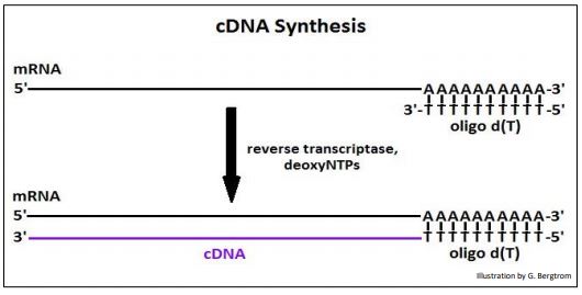
After heating to split up the cDNAs from the mRNAs, the cDNA is replicated to produce double-stranded, or (ds)cDNA, as illustrated below.
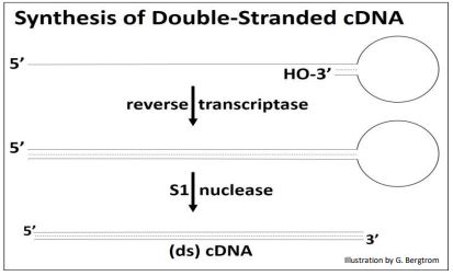
Synthesis of the 2nd cDNA strand is also catalyzed by reverse transcriptase! The enzyme recognizes DNA as well every bit RNA templates, and has the aforementioned v'-to-iii' DNA polymerizing activity every bit DNA polymerases. After 2nd cDNA strand synthesis, S1 nuclease (a unmarried-stranded endonuclease originally isolated from an East Asian fungus!) is added to open up the loop of the (ds) cDNA structure and trim the rest of the single-stranded DNA. What remains is the (ds) cDNA.
258 Isolate mRNA and Make cDNA
259 Reverse Transcriptase
B. Cloning cDNAs into Plasmid Vectors
To empathize cDNA cloning and other aspects of making recombinant Dna, nosotros need to talk a bit more than about the recombinant Dna tool kit. In addition to reverse transcriptase and S1 nuclease, other necessary enzymes in the 'kit' include restriction endonucleases (restriction enzymes) and Deoxyribonucleic acid ligase. The natural office of restriction enzymes in bacteria is to recognize specific restriction site sequences in phage DNA (most often palindromic Deoxyribonucleic acid sequences), hydrolyze information technology and thus avert infection.
Brake enzymes that make a scissors cut through the two strands of the double helix leaves blunt ends. Brake enzymes that make a staggered cutting on each strand at their restriction site leave behind complementary ('gluey') ends (beneath).

If you mix two of double-stranded DNA fragments with the same sticky ends from different sources (east.k., different species), they will form H-bonds at their complementary ends, making information technology easy to recombine plasmid DNA with (ds)cDNA, that have the same complementary 'gluey ends'. Using the language of recombinant DNA technologies, let's look at how plasmid vectors and cDNAs can be made to recombine.
1. Preparing Recombinant Plasmid Vectors Containing cDNA Inserts
Vectors are carrier DNAs engineered to recombine with foreign DNAs of interest. When a recombinant vector with its foreign Deoxyribonucleic acid insert gets into a host cell, it can replicate many copies of itself, enough in fact for easy isolation and study. cDNAs are typically inserted into plasmid vectors that are unremarkably purchased "off-the-shelf". They can exist cut with a restriction enzyme at a suitable location, leaving those gummy ends. On the other paw, it would not do to assimilate (ds)cDNA with restriction endonucleases since the goal is not to clone cDNA fragments, but entire cDNA molecules. Therefore, it will be necessary to adhere linkers to either end of the (ds)cDNAs. Plasmid DNAs and cDNA-linker constructs can then be digested with the same restriction enzyme to produce uniform 'glutinous ends'. Steps in the preparation of vector and (ds)cDNA for recombination are shown below.
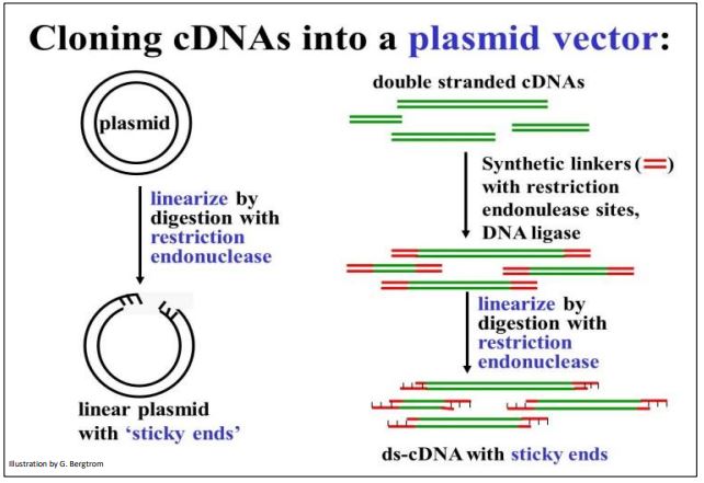
To prepare for recombination, a plasmid vector is digested with a restriction enzyme to open up the Dna circle. To take uniform viscid ends, double-stranded cDNAs to be inserted are mixed with linkers and DNA ligase to put a linker Deoxyribonucleic acid at both ends of the (ds) cDNA. Deoxyribonucleic acid ligase is another tool in the recombinant Deoxyribonucleic acid toolkit. Linkers are short, synthetic double-stranded DNA oligomers containing restriction sites recognized and cutting by the same brake enzyme as the plasmid. One time the linkers are fastened to the ends of the plasmid DNAs, they are digested with the advisable restriction enzyme. This leaves both the (ds)cDNAs and the plasmid vectors with complementary gluey ends.
260 Brake Enzymes and Recombinant Dna
2. Recombining Plasmids and cDNA Inserts and Transforming Host Cells
The next step is to mix the cut plasmids with the digested linker-cDNAs in only the correct proportions and then that the most of the cDNA (linker) ends volition anneal (class Hbonds) with the most of the mucilaginous plasmid ends. Adding DNA ligase to the plasmid/linker-cDNA mixture forms phosphodiester bonds between plasmid and cDNA insert, completing the recombinant circle of DNA, as shown beneath.
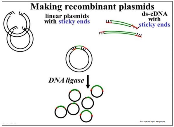
In early cloning experiments, an important consideration was to generate plasmids with merely one copy of a given cDNA insert, rather than lots of re-ligated plasmids with no inserts or lots of plasmids with multiple inserts. Using betterengineered vector and linker combinations, this issue became less important.
261 Recombine a cDNA Insert with a Plasmid Vector
3. Transforming Host Cells with Recombinant Plasmids
The recombinant DNA molecules are at present set up for 'cloning'. They are added to E. coli (sometimes other host cells) made permeable so that they can be hands transformed. Recall that transformation every bit divers by Griffith is bacterial uptake of strange DNA leading to a genetic change. The transforming principle in cloning is the recombinant plasmid! The transformation step is shown below.
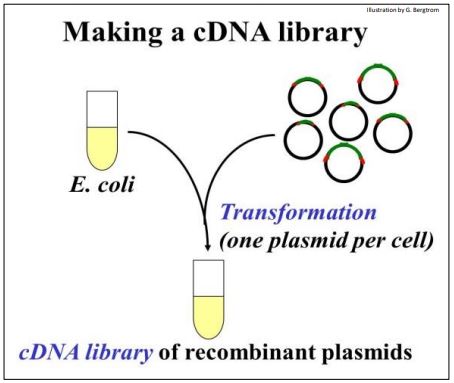
The tube total of transformed cells is the cDNA Library.
262 Making the cDNA Library.
After all these treatments, not all plasmid molecules in the mix are recombinant; some cells in the mix haven't even taken up a plasmid. So when the recombinant cells are plated on agar, how exercise you tell which of the colonies that grow came from cells that took upward a recombinant plasmid? Both the host strain of E. coli and plasmid vectors used these days were further engineered to solve this trouble. One such plasmid vector carries an antibiotic resistance gene. In this instance, ampicillin-sensitive cells would be transformed with recombinant plasmids containing the resistance cistron. When these cells are plated on media containing ampicillin (a form of penicillin), they grow, as illustrated below.
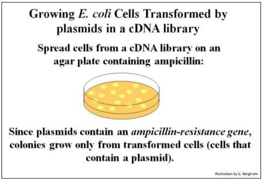
Untransformed cells (cells that failed to accept upwards a plasmid) lack the ampicillin resistance gene and thus, do not grow on ampicillin-medium. Only, there is still a question. How can you tell whether the cells that grew were transformed by a recombinant plasmid containing a cDNA insert? It is possible that some of the transformants comprise only non-recombinant plasmids that still have the ampicillin resistance gene!
To address this question, plasmids were further engineered with a streptomycin resistance gene. Only this antibody resistance gene was as well engineered to contain restriction enzyme sites in the middle of the gene. Thus, inserting a cDNA in this plasmid would disrupt and inactivate the gene. Here is how this second fleck of genetic technology enabled growth just of cells transformed with a recombinant plasmid containing a cDNA insert. Nosotros can tell transformants containing recombinant plasmids apart from those containing non-recombinant plasmids by the technique of replica plating shown (illustrated beneath).
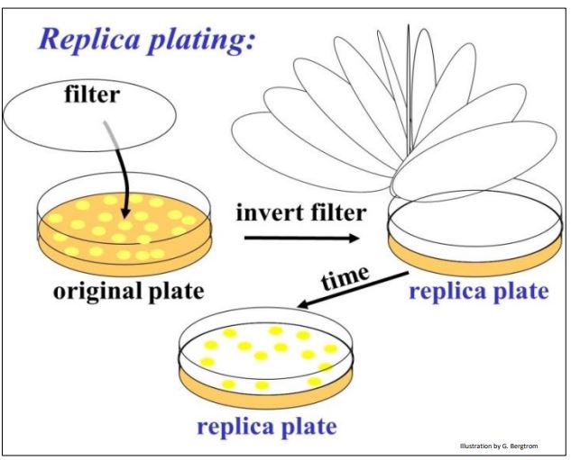
After colonies grow on the ampicillin agar plate, lay a filter over the plate. The filter will choice up a few cells from each colony, in effect becoming a replica (mirror image) of the colonies on the plate. Place the replica filter on a new agar plate containing streptomycin; the new colonies that grow on the filter must be streptomycin-resistant, containing only non-recombinant plasmids. Colonies containing recombinant plasmids, those that did not grow in streptomycin are easily identified on the original ampicillin agar plate. In practice, highly efficient recombination and transformation procedures typically reveal very streptomycinresistant cells (i.due east., colonies) after replica plating. In this instance, ampicillin-resistant cells constitute a adept cDNA library, ready for screening.
263 Making a Replica Plate Filter
4. Identifying Colonies Containing Plasmids with Inserts of Interest
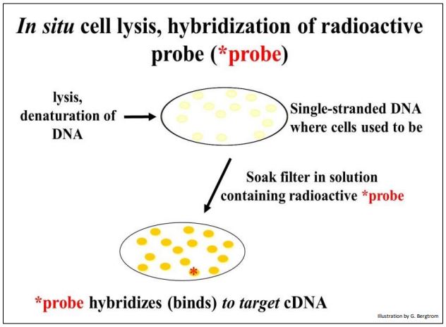
The side by side pace is to screen the colonies from the cDNA library for those containing the specific cDNA that y'all're after. This typically begins preparing multiple replica filters like the one above. Remember, these filters are replicas of bacterial cells containing recombinant plasmids that grow on ampicillin just non streptomycin. The number of replica filters that must exist screened tin be calculated from assumptions and formulas for estimating how many colonies must be screened to represent an entire transcriptome (i.e., the number of different mRNAs in the original cellular mRNA source). Once the requisite number of replica filters are made, they are subjected to in situ lysis to disrupt cell walls and membranes. The result is that the jail cell contents are released and the Dna is denatured (i.e., becomes single-stranded). The DNA and so adheres to the filter in identify (in situ, where the colonies were). The result of in situ lysis is a filter with faint traces of the original colony (below).
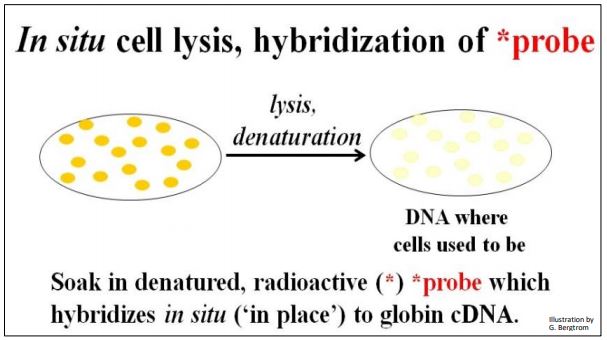
Side by side, a molecular probe is used to identify DNA containing the sequence of interest. The probe is ofttimes a synthetic oligonucleotide whose sequence was inferred from known amino acid sequences. These oligonucleotides are made radioactive and placed in a bag with the filter(southward). DNA from cells that contained recombinant plasmids with a cDNA of interest will demark the complementary probe. The results of in situ lysis and hybridization of a radioactive probe to a replica filter are shown below.
264 Probing a Replica Plate Filter
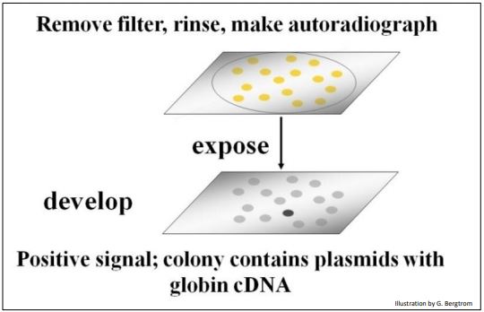
The filters are rinsed to remove un-jump radioactive oligomer probe, and then placed on X-ray film. After a menstruum of exposure, the film is developed. Blackness spots will class on the film from radioactive exposure, creating an autoradiograph of the filter. The black spots in the autoradiograph correspond to colonies on a filter that contain a recombinant plasmid with your target cDNA sequence (below).
In one case a positive clone is identified on the motion-picture show, the respective recombinant colony is located on the original plate. This colony is grown up in a liquid culture and the plasmid Deoxyribonucleic acid is isolated. At that point, the cloned plasmid DNA can be sequenced and the amino acid sequence encoded by its cDNA can be inferred from the genetic lawmaking dictionary to verify that the cDNA insert in fact encodes the protein of interest. Once verified equally the sequence of interest, a cloned plasmid cDNA can exist fabricated radioactive or fluorescent, and used to
- probe for the genes from which they originated.
- identify and quantitate the mRNA even locate the transcripts in the cells.
- quantitatively measure amounts of specific mRNAs.
Isolated plasmid cDNAs can fifty-fifty be expressed in suitable cells to make the encoded poly peptide. These days, diabetics no longer receive sus scrofa insulin, simply go synthetic homo insulin homo made from expressed homo cDNAs. Moreover, while the introduction of the polymerase chain reaction (PCR, see beneath) has superseded some uses of cDNAs, they nevertheless play a role in genome-level and transcriptome-level studies.
265 Pick a Clone From a Replica Filter and Play With It!

Why Are Filters Overlaid With Xã¢â‚¬â€˜ray Film When Screening A Cdna Library?,
Source: https://bio.libretexts.org/Courses/Ohio_State_University/Ohio_State_University_SP21:_Molecular_Genetics_4606_%28Chamberlin%29/10:_Amplifying_and_Manipulating_DNA_Fragments/10.07:_Make_and_Screen_a_cDNA_Library
Posted by: wisehumpertle.blogspot.com
0 Response to "Why Are Filters Overlaid With Xã¢â‚¬â€˜ray Film When Screening A Cdna Library?"
Post a Comment